Iliotibial Band Syndrome
Anatomy, Prevention & Treatment

Pain in the outer part of the knee can mean several things that could be a potential cause, but it migh be worth considering iliotibial band syndrome (ITBS).
ITBS in runners is a leading cause of lateral knee pain with accounting to
approximately 12% of all running related injuries (9, 4). It is the second leading cause
of overall knee pain and a non-traumatic overuse injury thought to be
caused by repeated flexion and extension of the knee which can irritate the
structures around the joint. The constant friction of the ITB repeatedly sliding forward and backward over the lateral femoral epicondyle
(figure 1) can be the cause of local inflammation of the ITB or outer knee (10, 7).
Other causes will be explained below which consider the extrinsic and intrinsic factors that can cause ITBS.
The Iliotibial Band and Anatomy
The ITB has been described as a large thick band of deep fascia and is a continuation of the fascia arising from the tensor fascia lata (TFL), gluteus maximus and gluteus medius muscles (figure 1) (1).
The connective tissue of the ITB assists stance stability and resists large varus torques at the knee and could be why this region becomes tight in individuals with a varus tibiofemoral strain (2). Iliotibial band syndrome occasionally becomes inflamed at its proximal origin and can cause referred hip pain.
The muscle is linked to the femur via strands of dense, regular fibrous connective tissue (3). After it courses between the biceps femoris and vastus lateralis, the ITB attaches to the lateral femoral condyle and sends lateral retinacular fibers to the patella (11).
The iliotibial band has two significant attachments including the lateral epicondyle, Gerdy's tubercle (at the anterolateral tibia) and the fibular head (figure 2).
The iliotibial band attachments include the distal edge of the femur at the upper edge of the lateral epicondyle which has a layer of adipose tissue underneath (2). The adipose tissue contains pacinian corpuscles which is highly vascular and may be the site of inflammation that causes pain during compression (2). Interestingly, certain movements can 'tug' at the Gerdy's tubercle attachment when the muscle is tensed, especially during internal rotation of the tibia as the knee bends during the weight-acceptance phase of gait (2).
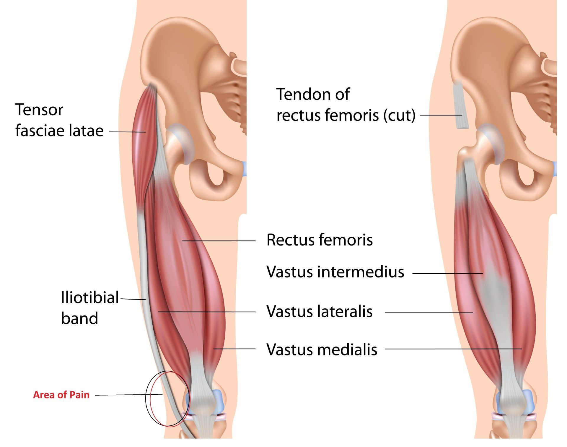
Figure 1
Intrinsic Contributing Factors To ITBS
Various intrinsic factors can attribute to ITBS and the following are signs that your therapist will be looking out for during a physical examination:
- Femoral external rotation during stance and gait cycle.
- Excessive hip adduction.
- Knee internal rotation.
- Leg length descrepencies.
- Increased foot inversion.
- Reduced Hamstring strength.
- Increased knee flexion at heel strike.
- Reduced hip abductor muscle strength.
- Weak hip abductors which can lead to increased hip adduction during stance phase of gait (7).
- Knee varus (bowlegs) or valgus (knock-knee) strain (figure 2).
- Shortenend and strengthened tensor fascia lata (postural muscle) which clinically tends to create increased flexion at the hip during stance phase and tends to increase hip internal rotation.

Figure 2
Extrinsic Contributing Factors
These include:
- Excessive running; greater than normal weekly mileage
- Downhill running as the reduced knee flexion at foot strike increases friction between the ITB and lateral epicondyle.
Abnormal Walking Biomechanics
Biomechanical studies in runners with ITBS show that the posterior edge of the ITB can impinge against the lateral femoral epicondyle of the femur just after foot strike in the gait cycle (6). This can produce irritation and a subsequent inflammatory reaction, especially in the region beneath the posterior fibers of the ITB which are felt to be tighter against the lateral femoral condyle than the anterior fibers.
The symptoms usually occur after a reproducible time or distance and consist of a sharp pain or burning on the lateral aspect of the knee. Occasionally, there will be swelling and thickening of the tissue where the band moves over the lateral femoral condyle (6).
Assessment
The Ober Test
The Ober test is the most commonly used to assess tightness of the iliotibial band which is performed via:
The examiner stands behind a side lying patient and stabilises the pelvis with one hand. The superior thigh is then moved into maximal abduction which is maintained in this position whilst moving the thigh into extension. The examiner lowers the limb into adduction until end range, or until the pelvis starts to tilt. The angle of hip joint adduction is considered to be a measurement of ITB length (7).
Angle of Knee Flexion During Stance Phase
Treating ITBS
Stretching
Wilhelm et al., (2017) investigate the iliotibial band tensor fascia lata complex (ITBTFLC) elongation response to a simulated clinical stretch (in-vitro) and whether a “stretch” to the region translates into a clinically meaningful tissue elongation. The study found that greater lengthening occurred in the proximal region of the ITBTFLC, suggesting that the proximal region (containing the TFL) is more likely to undergo elongation in response to a clinical stretch force when compared to the middle or distal regions (11).
However, the increased lengthening response in the proximal region of the ITB may be due to the presence of the TFL, which suggests that the ITB is incapable of elongating when applying a passive stretch (11).
Massage
A study by Chaudhry et al., (2008) found that large forces, outside the normal physiologic range of manual therapy, are required to produce even 1% compression and 1% shear in the tensor fascia lata (with 9075N or 925 kg)and in the plantar fascia (4515N, 460 kg). This indicates that it is virtually impossible to physically elongate the length of the tensor fascia or ITB (3). For this reason, I have learnt that it is more effective to apply massage and physical therapy to the quadratus lateralis and gluteal muscles which surround the ITB attachments and are often tight.
Correcting Biomechanics
Manual therapy might consist of correcting contributing intrinsic factors listed above and handing strengthening exercises to address weakness within gluteus medius and excessive hip adduction and knee internal rotation or excessive knee varus or valgus strain.
More information and exercises can be read in the attached here.
References
1.) AiKhaund, R., Flynn, S. H. (2005) Iliotibial Band Syndrome: A Common Source of Knee Pain, 71; 9: 1545-1550.
2.) Baker, R. L., Souza, R. B., Fredericson, M. (2011) Iliotibial Band Syndrome: Soft Tissue and Biomechanical Factors in Evaluation and Treatment, Clinical Review: Current Concepts, the American Academy of Physical Medicine and Rehabilitation; 3: 550-561.
3.) Chaudhry, H., Schleip, R., Ji, Z., B, B., M, M., F, T. (2008) Three-Dimensional Mathematical Model for Deformation of Human Fasciae in Manual Therapy, JAOA; 108: 8.
4.)Fredericson, M., Cookingham, C. L., Chaudhari, A. M., Dowdell, B. C., Oestreicher, N., Shirley A. Sahrmann, S. A. (2000)Hip Abductor Weakness in Distance Runners with Iliotibial Band Syndrome, ClinJ Sport Med; 3 (10): 169-175.
5.) Fredericson M., Guillet M., DeBenedictis L. (2000) Quick solutions for Movement Patterns. Iliotibial Band Syndrome. Phys Sportsmed, 28, 52-68.
6.) Fredericson, M., Wolf, C. (2005) Iliotibial Band Syndrome in Runners: Innovations in Treatment, Injury Clinic, Sports Med; 35 (5): 451-459.
7.) Lavine, R. (2010) Iliotibial Band Friction Syndrome, Humana Press; 3: 18-22.
8.) Miller, R. H., Lowry, J. L., Meardon, S. A., Gillette, J. C. (2007) Lower extremity Mechanics of Iliotibial Band Syndrome During An Exhaustive Run, Science Direct, Gait and Posture; 26: 407-413.
10.) van der Worp, M. P., van der Horst, N., de Wije, A., Backx, F. J., Nijhuis-van der Sanden, M. W. (2012) Iliotibial Bnd Syndrome in Runners: A Systematic Review, Sports Med, 1 42; 11: 969-92.